Table of contents of the article
ToggleBlackleg disease is a fungal disease that affects cabbage plants and leads to deterioration in production. In this article from the “WORLD OF PLANTS” website, we review the most important symptoms of infection and methods of prevention and control.
Cause of black leg disease
Plenodomus lingam scientific name
Type of disease: fungal
Families. It is found in cabbage, canola and cauliflower
Blackleg disease, also known as Phoma leg canker, is caused by two species of fungi, Leptosphaeria maculans and Leptosphaeria biglobosa. It spends its hibernation period on seeds, or on plant residues and crop residues that remain in the field. They then begin producing spores during the onset of warm, humid weather in the spring. These spores are dispersed by wind or rain to healthy plant parts, especially the lower leaves and the base of the stem. The germination of spores and the growth of fungi on plant tissues leads to the appearance of symptoms. If plants become infected, seedlings may die early in the season (seedling death). The fungus spreads from young leaves to the stem, where it grows to form ulcers at the junction between petioles and stems or around the crown. This in turn limits the transport of water and nutrients through the stem, and leads to death and stagnation. It is also an important disease on rapeseed and other crops of the cruciferous family (canola, turnip, broccoli, Brussels sprouts, cabbage).
Suitable conditions for blackleg disease on cabbage
The development of the disease is promoted by high air humidity (60-80%) and temperatures from 20 to 24 ° C. The incubation period of the disease lasts 5-8 days. During the vegetative period the fungus can produce 5-8 generations. Infected seeds and plant debris are a source of infection, and the infectious agent can be maintained for 2-3 years in the form of pycnidia.
Effect of blackleg disease on cabbage
The causative agent of black leg of cabbage seedlings is dangerous because it is very active. At the seedling stage, mass infection is often observed, due to which almost all seedlings die. Another danger is that the disease is difficult to treat. If it passes into a neglected form, it will not be possible to deal with it. Therefore, it is easier to prevent the development of the disease than to deal with the consequences. It is important to understand that the pest infects not only cabbage seedlings, but also other plants. In addition, it will definitely remain in the soil. Therefore, this soil must be disinfected or frozen. Otherwise, from year to year there will be significant crop losses.
Symptoms of black leg disease on cabbage
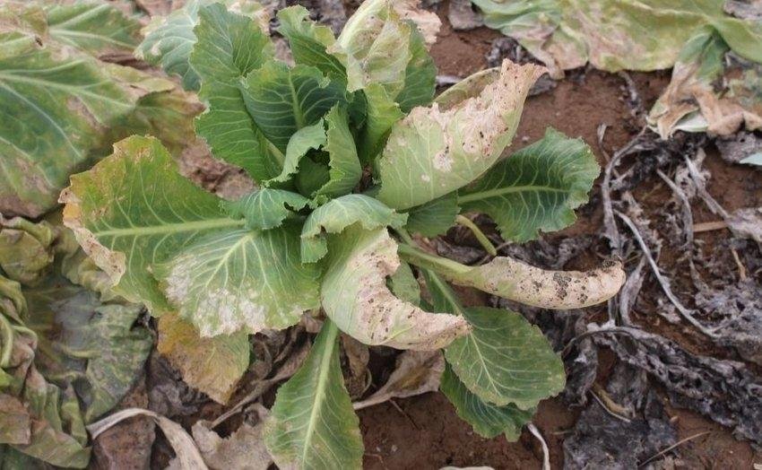
. Disseminated pale gray circular spots covered by either black dots, necrotic spots, or both appear on the leaves.
. Both types of spots are surrounded by a chlorotic (yellowish) halo.
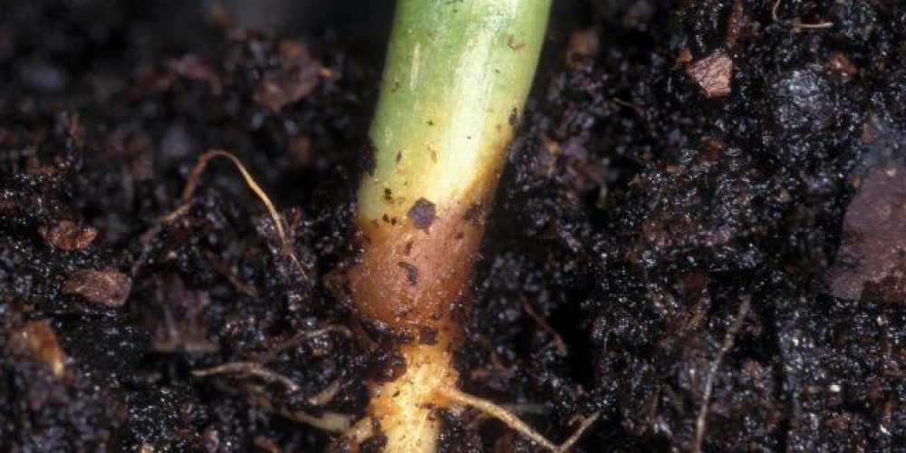
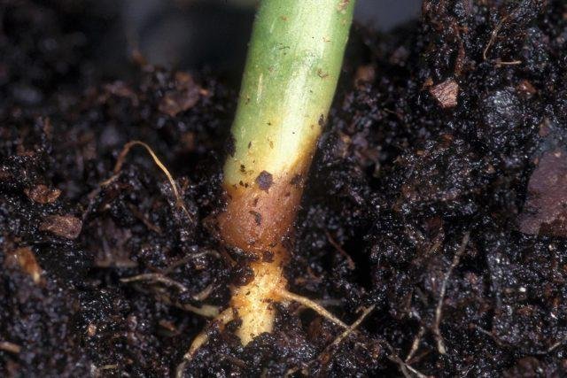
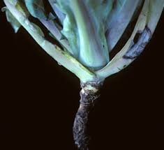
On the stems, gray spots appear that can develop into ulcers.
. As the stems grow, the cankers girdle and weaken the stem, leading to dormancy and death of the plant.
. The severity of symptoms varies greatly depending on the crop or variety, as well as the pathogen and prevailing environmental conditions. In any case, the main symptoms appear on the leaves and stems. Leaf spots include circular, pale gray spots dotted with black dots or with dark, dead, necrotic spots. Yellow discolouration in leaf veins or the areas surrounding the spots (chlorosis) is also common. Gray spots appear on the stems, which can range from small brown rectangular spots to ulcers that surround the entire stem. Black spots can also be observed on it. As these stems grow, the cankers girdle and weaken the stem, leading to premature maturation, dormancy, and death of the plant. The pods may also show symptoms in the form of brown spots with black margins, leading to premature maturation and seed infection.
Life cycle of blackleg disease on cabbage
The ascospores of the fungus are released after rainfall when temperatures range between 8-12°C (46-54°F). These spores can be spread by wind hundreds of meters (yards). Pycnidia can easily overwinter in tree debris, but because pycnidiospores are largely airborne, they are of secondary importance in initiating the first cycle of the disease
Ascospores germinate in the presence of free water at a temperature of 4-28°C (40-82°F). Penetration is through stomata. The pathogen may also be seed-borne. The seeds may be infected and/or infested with the pathogen. Infected seeds give rise to infected seedlings, but seed contamination levels are always very low. Primary infection usually occurs on the cotyledons or basal rosette leaves of the plant. Wet weather favors these primary infections
The fungus invades the intercellular spaces between the leaf septa and the epidermal layers. This asymptomatic vital stage is followed by mesophyll invasion with resulting cell death and the appearance of grey-green lesions. The hyphae continue to branch through the leaf tissue until they reach the leaf vein. The fungus then colonizes the cortex and/or wood parenchyma of the petiole. At the junction of the petiole and the stem, the fungus invades the cortex of the stem where it causes canker. At this point, stem resistance is expressed and determines the ability of the disease to move to the damaging stem canker stage. Stem cankers form when plants burst. They produce an upright stem from the rosette on which flowers are formed. Stem cankers develop most rapidly at 20-24°C (68-75°F) and are more serious under conditions of stress such as mechanical, insect or herbicide injury. Pycnidiospores (conidia) of Pycnidia under moist conditions in a mucilage, which is a watery and viscous solution. These spores are responsible for secondary courses of the disease, but Ascospores are the most important source of pus because they are more contagious and airborne. Penediospores are spread to new infection sites by rain droplets. Pycnidiospores germinate more slowly than ascospores and require more than 16 hours of continuous wetness at the optimum temperature range of 20-25°C (68-77°F). The minimum latency period from infection to production of new inoculum after infection with pycnidiospores is 13 days. Although secondary infections by pycnidiospores do occur, most losses are due to primary infections of leaves by ascospores that lead to basal stem cankers and eventual plant establishment.
Recommendations for blackleg disease on cabbage
We recommend applying organic control in the early stages of the disease or when the crop is close to harvest. In the more advanced stages of the disease, please use chemical control. It is not recommended to mix or use different products at the same time.
Organic control of blackleg disease on cabbage
There appear to be no biological control measures available to combat these diseases. Please contact us if you know of any.
Chemical control of blackleg disease on cabbage
Always follow an integrated approach with preventive measures together with biological treatment methods if available. Fungicides have very little effect once the fungus reaches the stem and the justification for using pesticides in fields is the expectation of high yields. Proticonazole can be used as a foliar spray. Effective seed treatment with prochloraz supplemented with thiram can reduce seedling infection caused by seed-borne Phosphorus.
Preventive measures for blackleg disease on cabbage
- The most important measure against this disease is the use of resistant varieties, if available for the chosen crop.
- And plan crop rotation with non-host plants.
- Practice deep plowing and bury crop residues after harvest.
- Surface tillage can also prevent fungi from reaching the leaves and lower stems.
In conclusion, we would like to note that we, at the world of plants website, offer you all the necessary services in the world of plants, we provide all farmers and those interested in plants with three main services::-
- Artificial intelligence consulting service to help you identify diseases that affect plants and how to deal with them.
- Blog about plants, plant diseases and care of various crops ... You are currently browsing one of her articles right now.
- An application that provides agricultural consultations to clients, as well as a service for imaging diseases and knowing their treatment for free – Click to download the Android version from Google Play Store، Click to download the IOS version from the Apple App Store.
References:
- Pillai VI, ed. 1988. Microorganisms. Plant disease factors. Kyiv: Naukova Dumka. 550 p. (in Russian)
- Hawksworth DL, Kirk PM, Sutton BC, Pegler DM 1995. Ainsworth & Bisby.s Dictionary of Fungi. Cap International. 616 p.
- Kvashnina E., Andreev N. 1935. Threat of cabbage production from poisoning. Na zashchitu urozhaya (OGIZ-Sel.khozgiz, VIZR), 3: 20-22. (in Russian)
- Mikhal.chuk NV 1984. Cabbage curd. Kartovel. I ovoshchi, 3: 26. (in Russian)
- Peresypkin VF 1969. Diseases of agricultural plants. Moscow: Kolos. 479 p. (in Russian)
- Sal.nikova AF 1952. Cabbage suffocation in Far Eastern conditions and control. Ph.D. Omsk. (in Russian)
- © Khlopunova LB
- Williams, PH 1992. Biology of Leptosphaeria maculans. Can. Y. Plant path. 14:30-35.
- Google, RK and GA Petrie. 1992 History, event, impact and control of blackleg of rapeseed. Can. Y. Plant path. 14: 36-45.
- Hall, R. 1992. Epidemiology of blackleg of oilseed rape. Can. Y. Plant path. 14: 46-55.
- Reimer, S.R. and C.J. vandenBerg.1992. Resistance of oilseed plants to Brassica spp. For blackleg caused by the fungus Leptosphaeria maculans. Can. Y. Plant path. 14: 56-66.
- Soledad, M., C. Piedras, and J. Seguin-Schwartz. 1992. Blackleg mushrooms: phytotoxins and phytoalexins. Can. Y. Plant path. 14: 67-75.

